
FIGURE 3.
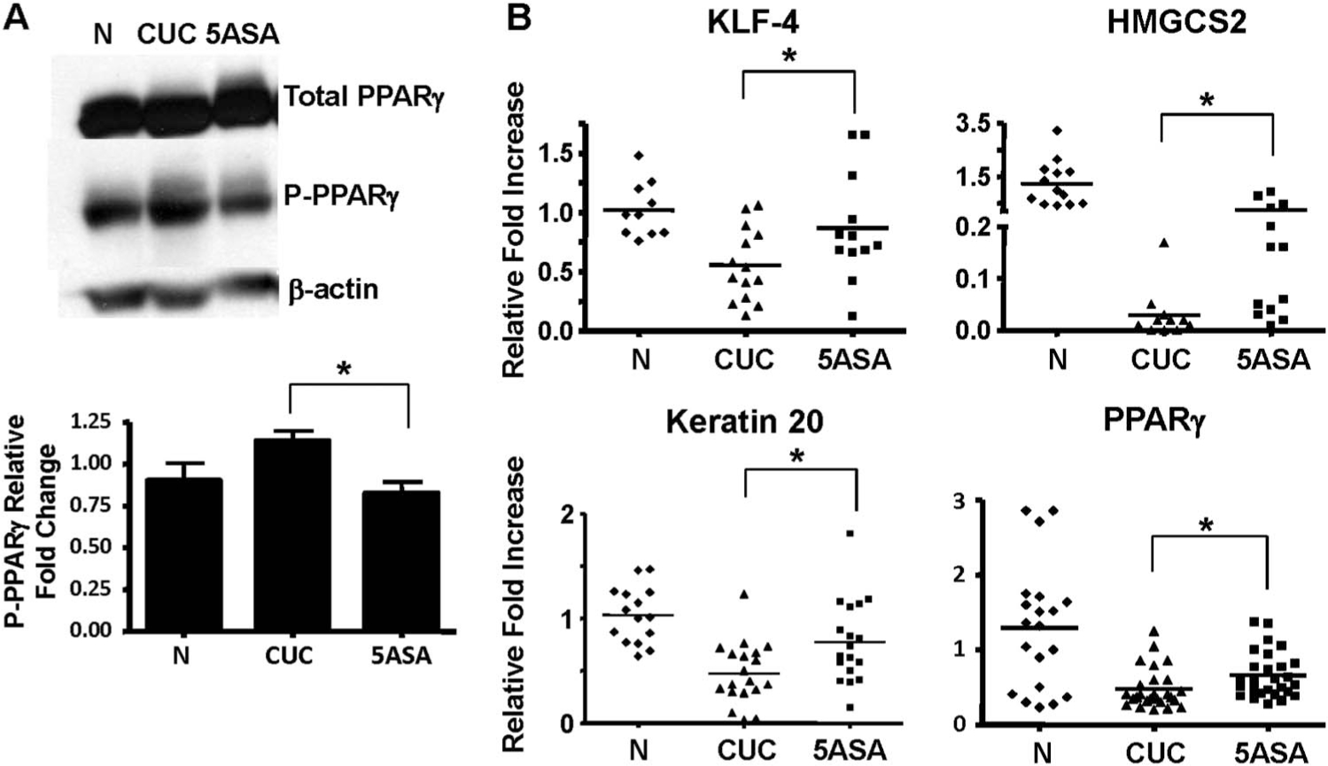
PPARγ activity is impaired in CUC and improved with 5-ASA. A, WB analysis of total PPARγ and transcriptionally inactive P-PPARγ from whole cell extracts from biopsies obtained from normal, active CUC, and 5-ASA–treated CUC patients (top). Densitometry of western blots performed on 5 individual patients per group indicates the decrease in P-PPARγ detection in CUC with 5-ASA compared to untreated patients (normalized to β-actin) is statistically significant. B, qRT-PCR of PPARγ target genes comparing normal and 5-ASA–untreated versus 5-ASA–treated CUC patients. * indicate significant changes in mRNA levels in the CUC patient groups. WB, Western blot.