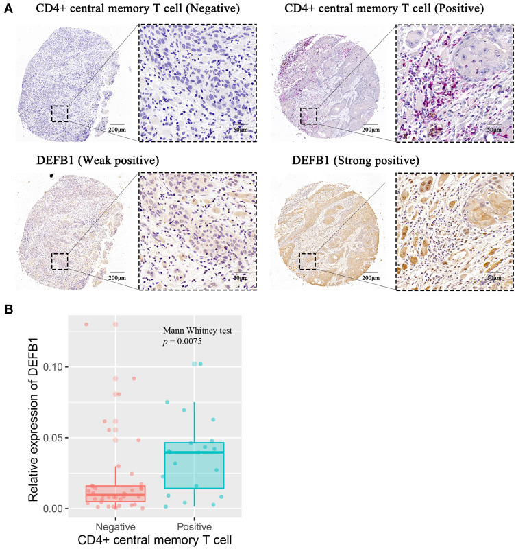
Figure 5.
The correlation between protein expression level of DEFB1 and infiltrating CD4+ central memory T cells in oral squamous cell carcinoma (OSCC) tissues. (A) Representative images of both high/low expression of DEFB1 and CD4+ central memory T cell at the same location in OSCC tissues. CD4+ central memory T cell was marked with both CD45RO+ (red) and CCR7+ (brown). (B) The correlation between DEFB1 expression and CD4+ central memory T cell infiltration in OSCC tissues. p value was calculated by Mann–Whitney test.

