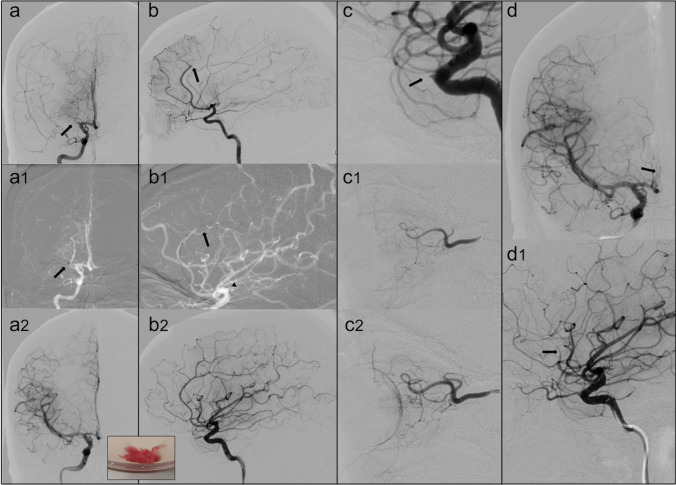
Fig. 2.
(a) The digital subtraction angiography (DSA) in frontal view confirms the occlusion of the right MCA (arrow). During the endovascular procedure (a1), the distal tip of the aspiration catheter (arrow) engages the thrombus with complete MCA recanalization (a2). In the box, the macroscopic aspect of the removed clot is shown. (b) DSA in lateral view reveals the occlusion of the A3 segment of the anterior cerebral artery (arrow). A combined technique with a stent retriever positioning thought thrombus in the pericallosal artery (arrow) and distal tip of aspiration catheter (arrowhead) at the origin of the vessel (b1) allows the ACA recanalization (b2). (c) DSA in lateral view: after the recanalization of intracranial vessels a right ophthalmic artery occlusion appeared (arrow). Micro-catheterization with intra-arterial alteplase infusion was performed (c1) with flow restoration (c2). (d) DSA in frontal and lateral view demonstrates the formation of further thrombi (arrows) with re-occlusion of the ACA

