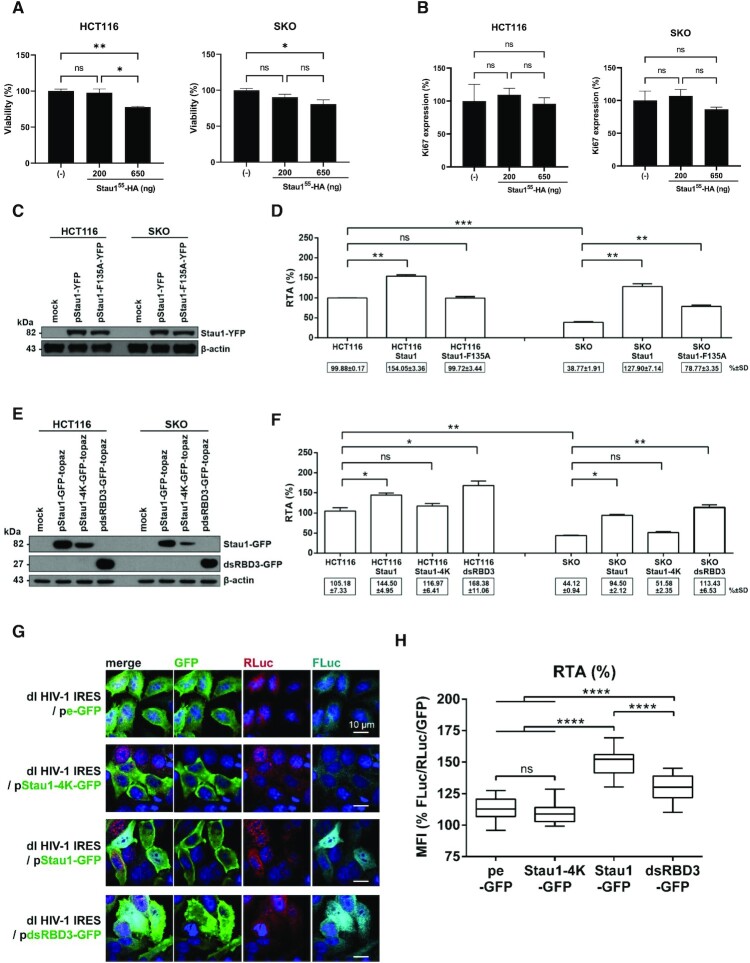
Figure 6.
Staufen1 and Staufen1-dsRBD3 rescues HIV-1 IRES activity in Staufen1 knockout HCT116 and HeLa cells. HCT116 or Staufen1 knockout HCT116 (SKO) cells were transfected, or not, with the Stau155-HA3 expression plasmid (200 or 650 ng), and cell viability (A) and cell proliferation (B) were analyzed by flow cytometry as indicated in Material and Methods. (C, D) The dl HIV-1 IRES plasmid (300 ng) was cotransfected, or not, with the Stau155-YFP expression construct was transfected into HCT116 or SKO cells. (C) The overexpression of the Stau155-YFP and Stau155-F135A-YFP in transfected HCT116 and SKO cells was confirmed by western blot using an anti-GFP antibody and β-actin as a loading control. (D) RLuc and FLuc activities were measured 24 h post-transfection, and results are presented as RTA. The RTA obtained for HCT116 cells transfected with dl HIV-1 IRES in the absence of Stau155-HA3, or the mutant protein was set to 100%. Statistical analysis was performed using an unpaired two-tailed t-test (ns, non-significant; * P ≤ 0.05; ** P ≤ 0.01; *** P ≤ 0.001; **** P ≤ 0.0001). (E, F) The dl HIV-1 IRES plasmid was cotransfected, or not, with the Stau155-GFP-topaz, mutant Stau155-4K-GFP-topaz or Stau155-dsRBD3-GFP-topaz expression construct into HCT116 or SKO cells. (E) The expression of Stau155-GFP-topaz, Stau155-4K-GFP-topaz, and Stau155-dsRBD3-GFP-topaz was confirmed by western blot using an anti-GFP antibody and β-actin as a loading control. (F) RLuc and FLuc activities were measured 24 h post-transfection, and results are presented as RTA. The RTA obtained for the HCT116 cells that did not overexpress Stau155-recombinant proteins or any of its domains was set to 100%. Statistical analysis was performed using an unpaired two-tailed t-test (ns, non-significant; * P ≤ 0.05; ** P ≤ 0.01; *** P ≤ 0.001; **** P ≤ 0.0001). (G, H) HeLa cells were cotransfected with the dl HIV-1 IRES plasmid together with the pe-GFP control plasmid, Stau155-4K-GFP-topaz, Stau155-GFP-topaz, or Stau155-dsRBD3-GFP-topaz expression constructs. (G) Immunofluorescence imaging demonstrating expression and localization of peGFP, Stau155-4K-GFP-topaz, Stau155-GFP-topaz, and Stau155-dsRBD3-GFP-topaz (green) in addition to that of RLuc (red) and Fluc (cyan) in HeLa cells cotransfected with the dl HIV IRES plasmid. (H) Graphical representation of results shown in panel (G), calculating [RTA/GFP] from imaging data using the mean fluorescence intensity (MFI) values obtained for FLuc, RLuc, and GFP. Statistical analysis was performed using a one-way ANOVA with Tukey post-test for multiple comparisons. ** P ≤ 0.01; **** P ≤ 0.0001.

