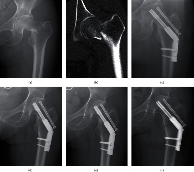
Figure 6.

Typical case of positive buttress reduction (female, 52-year-old). (a) Preoperative AP radiograph. (b) CT scan of the same patient. (c) Radiograph immediately after surgery showing positive buttress reduction. (d–f) Radiographs at 1 month, 3 months, and 12 months of follow-up: no complication occurred.
