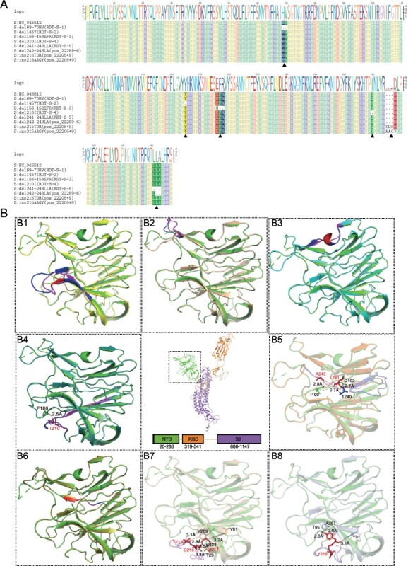
Figure 5.

Structural analysis of spike glycoprotein with recurrent indels. (A) The multiple sequences alignments display of RDT-S-1, RDT-S-2, RDT-S-3, RDT-S-4, RDT-S-5, pos_22289-6, pos_22205+9, pos_22206+9. (B) Tertiary structure of the recurrent indels of spike glycoprotein. B1–B8 are different S protein NTD indels tertiary structure align with template. (B1–B8) del69-70HV (yellow), del145Y (wheat), del156-158EFR (cyan), del210I (forest), del241-243LLA (orange), del242-243LA (splitpea), ins215TDR (light wheat), ins215AAGY (silver) and align with template PDB: 7CWU (green). Deletion or insertion area (red), amino acid change region (pink), corresponding normal area by amino acid change region (blue).
