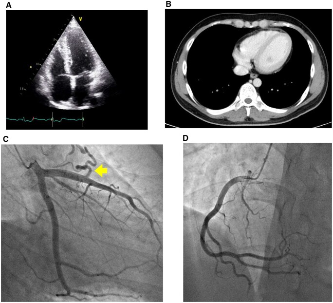
Figure 2.
Echocardiogram and thoracic computed tomography showed no pericardial effusion or ventricular wall thickening (A and B). Coronary angiography demonstrated no significant stenosis of the coronary arteries except for a coronary artery (left anterior descending) to pulmonary artery fistula (arrow) (C and D).

