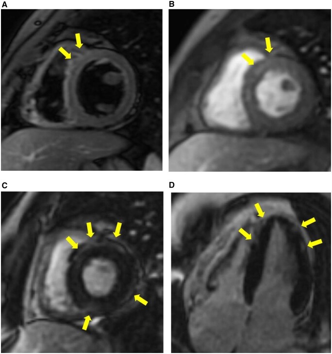
Figure 3.
Cardiac magnetic resonance images. Short T1 inversion recovery axis image acquired along the basal short-axis view demonstrates increased subepicardial and mesocardial signal intensity of the antero-septal myocardial segments (A). Dynamic contrast-enhanced sequence demonstrates slight elevated signals of the antero-septal myocardial segments (B). Fast imaging employing steady state acquisition sequences acquired along the basal short-axis view (C) and four-chamber view (D) demonstrate myocardial late gadolinium enhancement with epicardial predominance in the antero-septal, inferior and lateral walls of the basal segment and apex.

