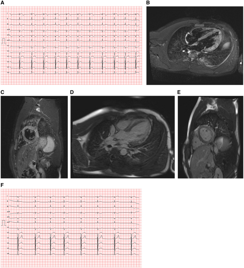
Figure 2.
(A) Case 2 electrocardiogram on presentation, showing ST-elevation in I, aVL, V4–V6, ST-depression in V1 and aVR. (B) Case 2 cardiac magnetic resonance imaging, T2-weighted stir sequence, four-chamber view, showing diffuse increased signal intensity in the anterolateral and apical segments. (C) Case 2 cardiac magnetic resonance imaging, T2-weighted stir sequence, midventricular short-axis view, showing an increased signal intensity in the mid inferolateral and anterolateral segments. (D) Case 2 cardiac magnetic resonance imaging, inverse-recovery late gadolinium enhancement sequence, four-chamber view, showing subepicardial late gadolinium enhancement of the anterolateral and apical segments. (E) Case 2 cardiac magnetic resonance imaging, inverse-recovery late gadolinium enhancement sequence, midventricular short-axis view, showing subepicardial late gadolinium enhancement of the mid inferolateral and anterolateral segments. (F) Case 2 electrocardiogram at Day 7, showing sinus rhythm, biphasic T waves in I, aVL, and ST-depression in III, aVR, and V1.

