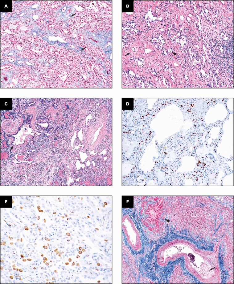
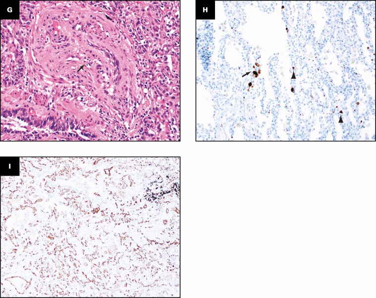
Figure 4.
Patient 2: Histopathologic findings in lung explant of the 33-year-old man with coronavirus disease 2019. The lungs show interstitial fibrosis varying from mild interstitial fibrosis (arrows, A) to more marked fibrosis (arrow, B) with collapsed alveoli (arrowhead, B). Peribronchiolar metaplasia is present (arrow, C). Immunostain for CD3 highlights the interstitial lymphocytic inflammation (D) and that for CD68 (E) highlights the numerous macrophages. The wall of the small airways has fibrosis (arrowhead, F), and the accompanying muscular artery has eccentric intimal fibrosis (arrow, F) that likely represents a healed thrombus. Recanalized thrombus is present in muscular artery (arrow,G). Immunostain for CD61 (H) shows microvascular thrombi (arrow, H) and platelets (arrowheads, H) in the parenchyma. Immunostain for CD34 shows increased interstitial capillary density (I). B, C, G, H&E; A, F, trichrome; E, CD68; H, CD61; I, CD34. A, B, D, ×10; C, F, ×4; E, ×40; G-I, ×20.


