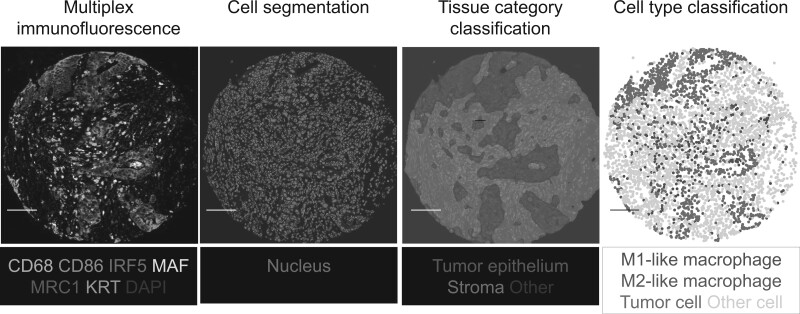
Figure 2.
Representative images of the quantification of macrophage counts and polarization in the colorectal cancer microenvironment using a customized multiplex immunofluorescence assay. The multiplex immunofluorescence images were processed with pathologist-supervised image analysis algorithms to perform cell segmentation, tissue category classification, and cell type classification. The scale bar is 100 µm. The details were described in our previous article (26).

