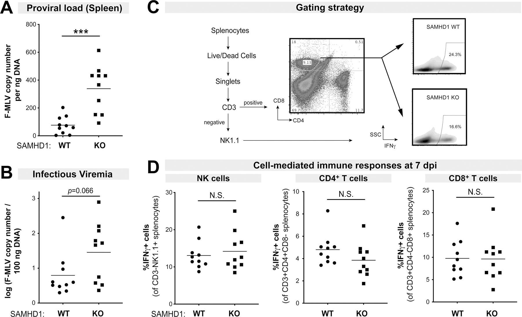
Figure 1.

Effects of SAMHD1 on FV infection and cell-mediated immune responses in the absence of LPS. B6 SAMHD1 WT (n=10) and KO mice (n=10) were infected with 104 spleen focus forming units of FV without LPS pre-treatment, and at 7 dpi, spleen and plasma were harvested. FV infection were evaluated for (A) Proviral loads by qPCR of spleen DNA and (B) infectious viremia by measuring viral DNA levels in target Mus dunni cells after 48 h following incubation with 5µl plasma. Splenocytes were subjected to flow cytometry as shown in the (C) gating strategy. The WT and KO panels were derived from samples in Figure 4C. (D) Splenocytes were evaluated for IFNγ expression in (left) NK cells; (middle) CD4+ T cells and (right) CD8+ T cells by flow cytometry. For all panels, each dot corresponds to a mouse. Data were combined from 2 independent mouse cohorts that included both male and female mice (Fig. S1A). Two-way comparisons were evaluated using a 2-tailed Student’s t-test. ***, p<0.001; NS, not significant.
