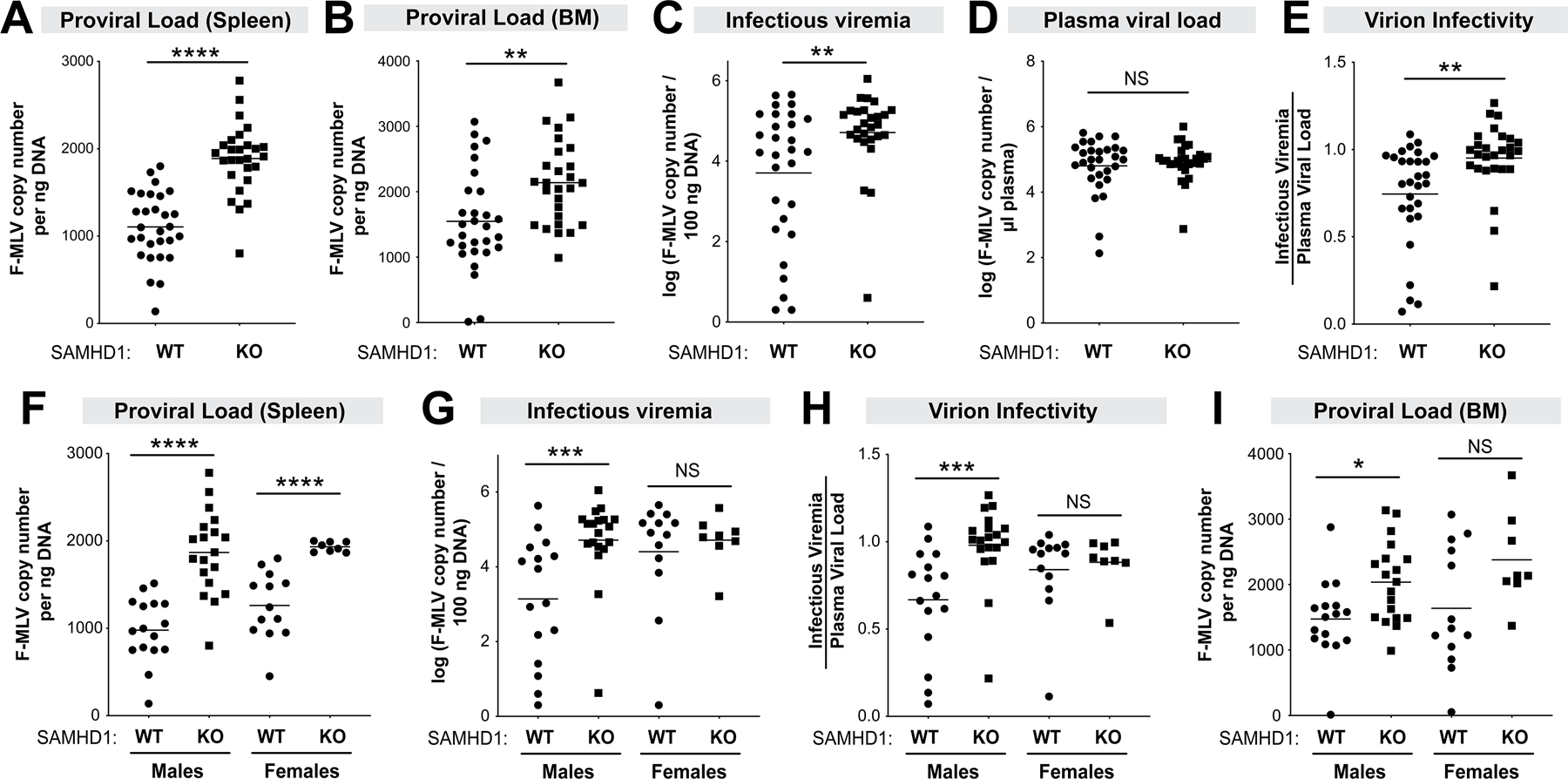
Figure 3.

SAMHD1 protects against FV infection in mice pre-treated with LPS. WT (n=29; 16 males, 13 females) and SAMHD1 KO (n=27; 19 males, 8 females) mice were infected with 104 SFFU of FV complex. Plasma, spleen and BM samples were harvested at 7 dpi to evaluate FV infection levels using different methods. Proviral DNA load in the (A) spleen and (B) bone marrow were measured by qPCR and normalized to ng of input DNA. (C) Infectious viremia were determined by co-incubating 5µl plasma with Mus dunni cells for 48 h and measuring F-MLV DNA copy numbers; (D) Plasma viral load was measured using F-MLV qPCR. (E) Virion infectivity was calculated by taking the ratio of log-transformed plasma infectious titer (C) and viral load (D). (Lower panels) Data from panels A, C, E and B were analyzed separately for males and females for (F) proviral load in the spleen, (G) infectious viremia, (H) virion infectivity) and (I) proviral load in the bone marrow, respectively. For all panels, each data point corresponds to a mouse. The data in the upper panels were pooled from 5 independent cohorts of WT and SAMHD1 KO mice. Each experiment consisted of WT (n=5–8) and SAMHD1 KO (n=3–8) mice. Log-transformed data were analyzed using a 2-tailed unpaired Student’s t-test. *, p<0.05; **, p<0.01; ***, p<0.001; ****, p<0.0001; NS, not significant (p>0.05).
