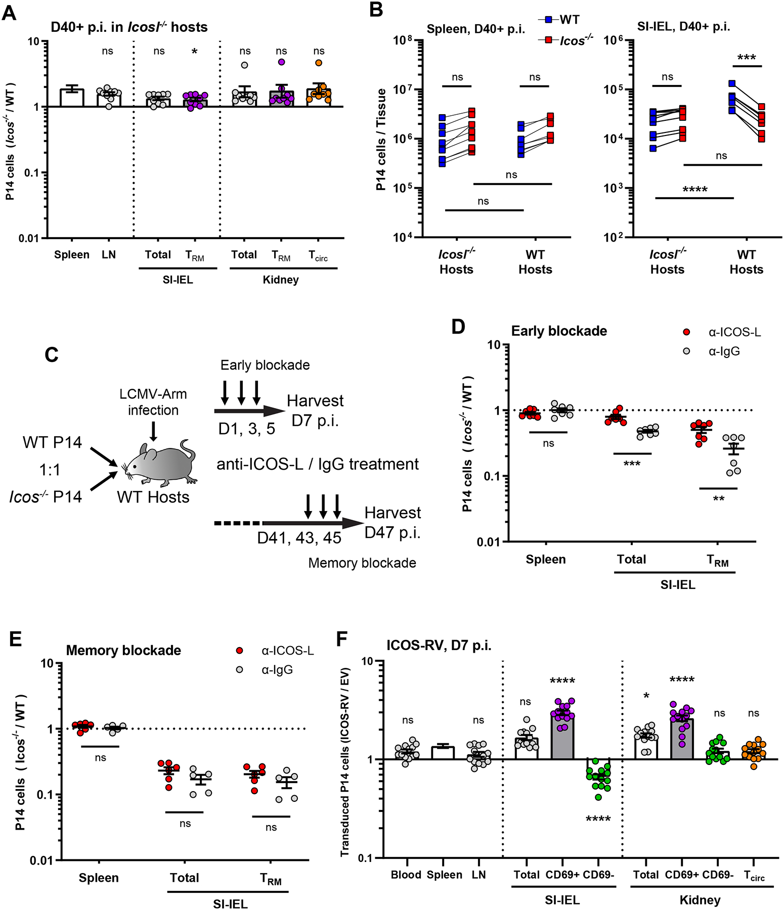
Figure 3. ICOS/ICOS-L interaction is essential for the optimal CD8+ Trm establishment.

(A) WT and Icos−/− P14 T cells were co-adoptively transferred into Icosl−/− recipient mice followed by LCMV infection. The ratio of transferred P14 T cells was determined in the indicated tissues ≥40 days later; Data are from 3 independent experiments with a total 9 recipient mice. (B) shows the absolute numbers of WT or Icos−/− donor P14 T cells in either WT or Icosl−/− recipient mice at 40+ days following LCMV infection, for the spleen and SI-IEL. Data are from 3 independent experiments with a total 8–9 recipient mice. (C–E) WT host mice received equal number of WT and Icos−/− P14 T cells were treated with ICOS-L blocking antibody (or isotype control) either early (D1–5) or late (D41–45) following LCMV infection, as illustrated in (C), and the ratio of donor populations determined at the indicated time points (D, E). Data are from 2 independent experiments with a total 5–7 recipient mice.
(F) Congenically distinct WT P14 T cells were transduced with retroviruses encoding ICOS (ICOS-RV) or an empty vector (EV) and co-transferred into WT hosts that were then infected with LCMV. At 7 days post infection, cells were isolated from the indicated tissues and the ratio between transduced cell populations was assessed. Data are from 3 independent experiments with a total 13 recipient mice.
Error bars represent mean ± SEM. Statistical significance was calculated with: Kruskal-Wallis test with multiple comparisons relative to spleen in (A), One-way ANOVA with multiple comparisons in (B), unpaired t-test in (D and E), and One-way ANOVA with multiple comparisons relative to spleen in (F): ns, not significant (p>0.05); * p<0.05; ** p<0.01; *** p<0.001; **** p<0.0001.
