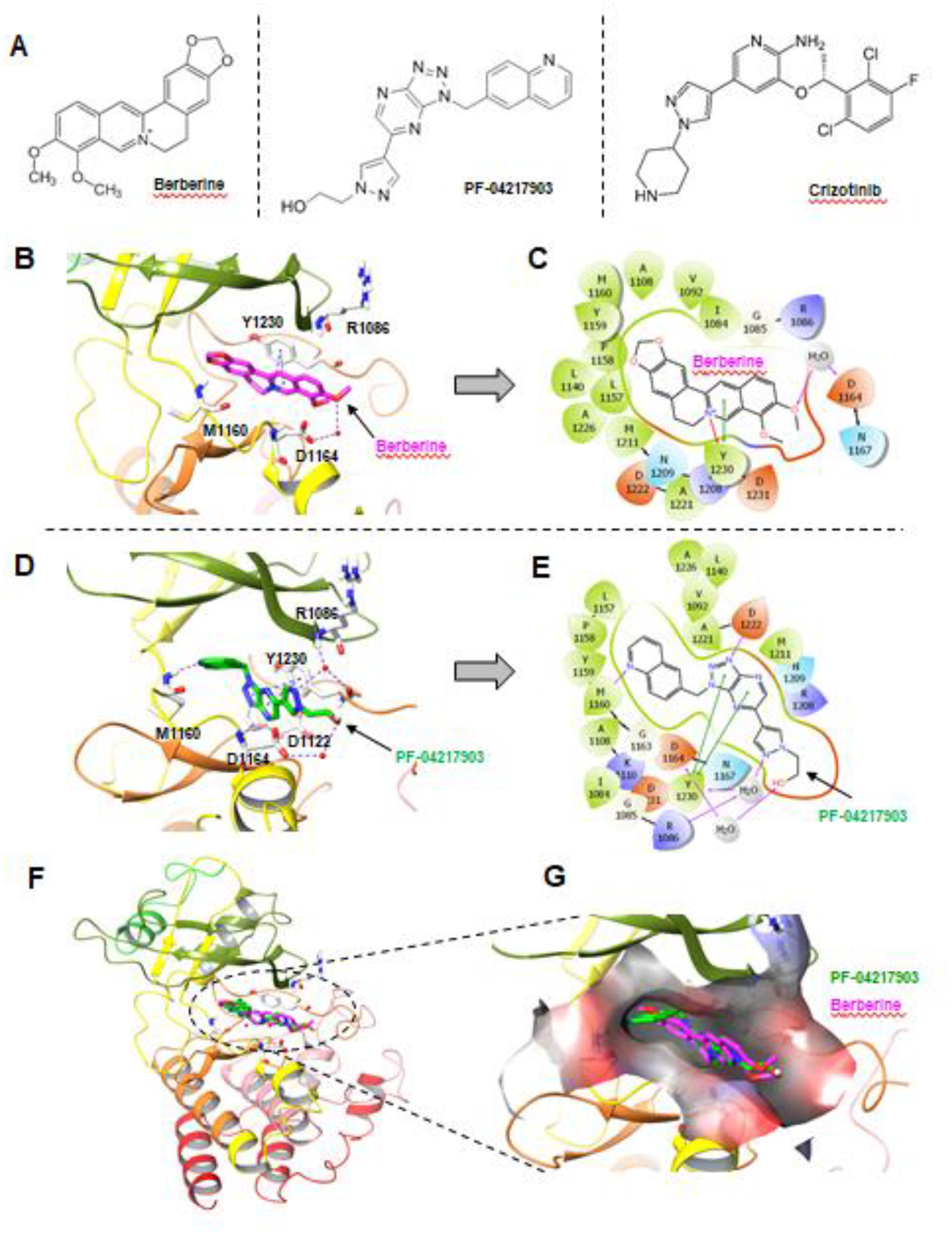
Fig. 6. Berberine can potentially bind to MET in a docking analysis.

A, Chemical structures of berberine and other known MET inhibitors. B, Berberine docking into MET kinase domain. H-bond: purple dashed line; π-π and π-cation: blue dashed lines; Berberine: magenta sticks; Water molecules: red spheres. C, Interaction diagram of Berberine docking pose. H-bond: purple line; π-π: green line; π-cation: red line. D, Binding site of the MET inhibitor, PF-04217903. H-bond: purple dashed line; π-π: blue dashed lines; PF-04217903: green sticks; Water molecules: red spheres. E, Interaction diagram of PF-04217903. F. Overlay of Berberine docking pose with MET protein co-crystal structures (PDB ID: 3ZXZ). Berberine: magenta; PF-04217903: green. G, Berberine docking pose and PF-04217903 overlay at the binding pocket of MET kinase domain in surface representation. Berberine: magenta; PF-04217903: green.
