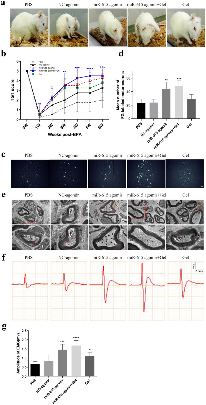
Fig. 3.
Co-transplantation of miR-615 agomir with gel improved the motor functional restoration of monoplegia right upper limb after avulsion/reimplantation. a Locomotion evaluation of animals in each group at sixth week after BPA-reimplantation. These pictures show a significant motor functional restoration that appeared on miR-615 agomir and miR-615 agomir+gel group, whereas the rats in other groups performed a little recovery. b Terzis grooming test was carried out weekly post BPA. Rats treated with miR-615 agomir+gel acquired the highest scores from the third week to sixth week after BPA reimplantation. c Fluorescence photomicrographs of FG-labeled motor neurons in ipsilateral C5–7 spinal segments. d Relative amounts of fluorogold-labeled motor neurons in ipsilateral C5–7 spinal cord. The number of fluorogold-labeled motor neurons in miR-615 agomir and miR-615 agomir+gel groups were more than PBS group. e Electron micrographs of musculocutaneous nerve. Distinct demyelination (red arrow) was observed in PBS, NC-agomir, and gel groups, yet the axons treated with miR-615 agomir with or without gel were more intact. f Electrophysiological activity of avulsed right forelimbs in each group at 6-week endpoint. g The amplitude of electromyography appears larger in the miR-615 agomir+gel group compared to PBS group, indicating better recovery of neuromuscular activity. Data are presented as the mean ± SD (one-way analysis of variance followed by the least significant difference post hoc test). *p < 0.05, **p < 0.01, ***p < 0.001, vs. PBS group. Scale bar (c) = 50 μm, Scale bar (e—upper row) = 1 μm, scale bar (e—lower row) = 0.5 μm

