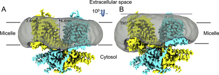
Fig. 5. Interaction between prestin and the surrounding micelle.
A Cryo-EM map for the gerbil prestin structure (colored by subunit in cyan and yellow, respectively) showing the surrounding micelle as a gray transparent surface. The dashed line (in navy) follows the micelle’s boundaries. The micelle belt around the transmembrane domain of prestin is locally distorted (thinned) at the cytosolic side close to TM6 and TM12 helices. The approximate thickness of the micelle in these regions is indicated by black arrows. The transmembrane helices 6, 7, and 12 are labeled. B As in (A) but tilted by ~10° to better illustrate the micelle’s distortions at the cytosolic side. See also Supplementary Fig. 9.

