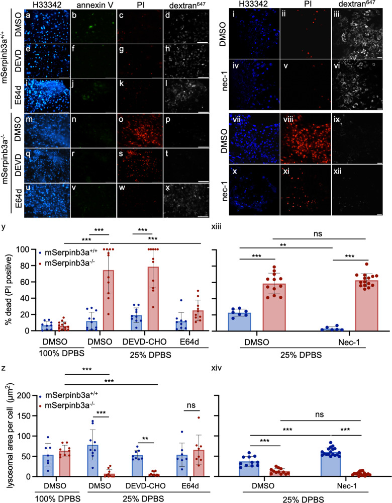
Fig. 2. LMP and lysosomal cysteine peptidases, but not executioner caspases or RIPK1, induced lysoptosis-like death in mSerpinb3a−/− FIECs.
a-x Representative confocal fluorescence images of mSerpinb3a+/+ (a-l) or mSerpinb3a−/− (m-x) FIECs pretreated with diluent (DMSO; a-d; m-p), 2 µm DEVD-CHO (e-h; q-t) or 2 µm E64d (i-l; u-x) and then incubated in 25% DPBS for 1 h (scale bar = 40 µm). FIECs were stained with Hoescht 33342 (H33342; blue), annexin V-FITC (annexin V, green), propidium iodide (PI, red), and Alexafluor647 conjugated dextran (dextran647, white). y, z Quantification of dead cells (PI-positive; y) or lysosomal area (dextran647 staining; z) from different representative experiments (n = 3) over multiple fields (n > 5). i-xii Confocal maximum intensity projections of fluorescently labeled lysosomes from mSerpinb3a+/+ (i–vi) or mSerpinb3a−/− (vii–xii) FIECs (scale bar = 25 µm). Cultures were pretreated with DMSO (i-iii; vii-ix) or nec-1 (iv-vi; x-xii) for 1 h prior to incubation in 25% DPBS. Cell numbers, cell death, and lysosomes were quantitated using H33342 (blue), PI (red), and dextran647 (white), respectively. Representative images are from the same experiment, which was repeated three times. xii, xiv Quantification of PI positive (xii) or lysosomal (xiv) staining from multiple fields (n ≥ 6). The means ± SD of a representative experiment were compared using a two-way ANOVA with Tukey’s multiple comparisons (**P < 0.01, ***P < 0.001). Original data files can be found at https://data.mendeley.com/datasets/scgbb3s333/1.

