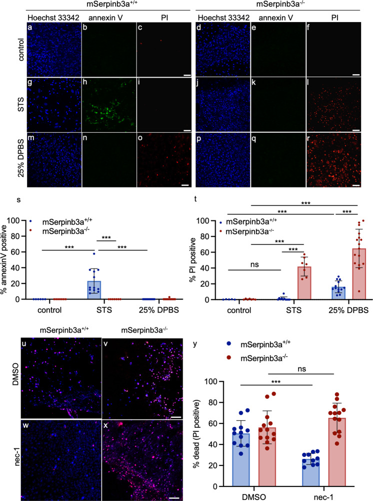
Fig. 3. Apoptosis or necroptosis inducers preferentially triggered lysoptosis-like cell death in mSerpinb3a−/− FIECs.
a–r To assess apoptosis, mSerpinb3a+/+ or mSerpinb3a−/− FIECs were treated with 100% PBS (a–f), 1 µm staurosporine (STS) for 16 h (g–l), or 25% PBS for 2 h (m–r). Representative confocal fluorescence maximum intensity projections were of cells stained with Hoechst 33342 (blue), annexin V-FITC (green), and PI (red; scale bars = 25 µm). s–t Quantification of annexin V (s) and PI (t) staining in multiple fields (n ≥ 5) from a representative of three experiments using mSerpinb3a+/+ (blue) or mSerpinb3a−/− (red) FIECs treated with 100% PBS (control), 1 µm STS for 16 h or 25% PBS for 2 h. u–x To induce necroptosis, mSerpinb3a+/+ (u, w), or mSerpinb3a−/− (v, x) FIECs were pretreated with either DMSO or 5 µm nec-1 and then incubated with 1 µm STS with 10 µm z-VAD-fmk for 16 h. Representative merged confocal fluorescence maximum intensity projections were of cells stained with H33342 (blue) and PI (red, scale bar = 25 µm). Dually labeled nuclei indicated dead cells and were depicted as magenta. y Quantification of PI staining in multiple fields from a representative of three experiments of mSerpinb3a+/+ or mSerpinb3a−/− FIECS pretreated with DMSO or 5 µm nec-1 and induced for necroptosis by treatment with 10 µm STS with 1 µm z-VAD-fmk for 16 h. For data in s, t, and y, the means ± SD were compared using a two-tailed t-test (***P < 0.001, **P < 0.01). Original data files can be found https://data.mendeley.com/datasets/4gfrf8xkyf/1.

