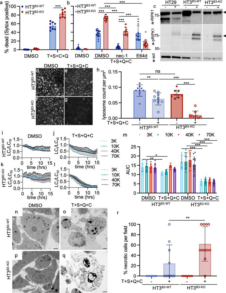
Fig. 9. Tumor epithelial cell lines null for SERPINB3 undergo lysoptosis-like death under conditions that induce RIP1K-dependent necroptosis.
a Percent dead ((# Sytox positive nuclei/# of blue nuclei) × 100) of HT3B3-WT or HT3B3-KO cells 12 h after incubation with DMSO or 10 ng/µL human TNFα + 0.1 µm BV6 SMAC mimetic + 0.5 µm qVD-OPh + 1 µm cyclohexamide (T + S + Q + C). b Percent dead (calculated as in panel a) of HT3B3-WT or HT3B3-KO cells incubated with DMSO, 50 µm nec-1 or 10 µm E64d for 1 h prior to exposure to T + S + Q + C for 8 h. c Immunoblot analysis of RIP kinase 1 (α-RIPK1), phosphorylated RIP kinase 1 (α-pRIPK1), and actin (α-act) in HT29 (control cell line), HT3B3-WT, and HT3B3-KO treated with DMSO (−) or T + S + Q + C (+) for 12 h (black arrowheads denote bands for RIPK1 and pRIPK1 based on molecular mass). d–g Representative confocal images (scale bar = 25 µm) of HT3B3-WT or HT3B3-KO cells incubated with DMSO or T + S + Q + C and stained with Lysotracker Deep Red (white). h Quantification of the lysosomal count per cellular µm2 for the experiment in d–g (≥9 fields, compared using a one-way ANOVA with Tukey’s multiple comparisons). i–l To quantitate lysosomal content, HT3B3-WT or HT3B3-KO cells were incubated with 3 kDa Cascade blue, 10 kDa Alexa488, 40 kDa TMR, and 70 kDa Texas red labeled dextrans prior to exposure to either DMSO or T + S + Q + C and imaged using live-cell resonance scanning confocal microscopy (≥20 z-planes, ≥10 fields). The number of lysosomes at each time point (LCt) were normalized to the lysosome count at time zero (LCt0). m AUC for each dextran over time for an experiment in i–l (two-way ANOVA with Tukey’s multiple comparisons test). n–q Representative TEM images (scale bars = 2 µm) of HT3B3-WT or HT3B3-KO cells treated with DMSO or T + S + Q + C. r Quantification of the number of necrotic cells per field (≥10 fields) for the experiment in n–q. Unless otherwise noted, a representative of ≥3 replicates is shown, and the presented means ± SD were compared using a two-tailed t-test (ns not significant, ***P < 0.001, **P < 0.01, *P < 0.05). Original imaging data can be found at https://data.mendeley.com/datasets/gvgnvkw4ct/1. Uncropped immunoblots can be found in Supplementary Fig. 21.

