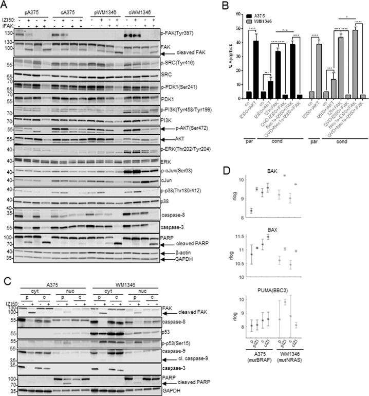
Fig. 6. Nuclear FAK prevents p53 activation in response to IZI50 treatment.
A Parental (p) and IZI5-conditioned (c) A375 and WM1346 cells were stimulated with IZI50, 5 µM defactinib (iFAK), or both. After 24 h the phosphorylation status of FAK, SRC, PDK1, PI3K, AKT, ERK, cJUN, and p38, as well as processing of caspase-3 and PARP was monitored by Western-blot analysis with β-actin and GAPDH as loading controls. One representative out of three independently performed experiments is shown. B Parental (p) A375 and WM1346 cells were stimulated with IZI50. IZI5-conditioned (c) cells were treated with either 5 µM QVD, 15 µM Nec1s, or both for 1 h prior to combined FAK inhibitor (iFAK) defactinib and IZI50 stimulation. After 24 h apoptosis induction was assessed using a CDDE (n = 3: *p ≤ 0.05; ***p ≤ 0.001; ****p ≤ 0.0001; n.s. = not significant). C Parental (p) and IZI5-conditioned (c) A375 and WM1346 cells were stimulated with IZI50. After 24 h the status of FAK, p53, p-p53(Ser15), caspase-8, caspase-3, caspase-9 and PARP, respectively, was determined in cytosolic (cyt) and nuclear (nuc) protein fractions by Western-blot analysis, with GAPDH as loading control. One representative out of three independently performed experiments is shown. D Mean expression (rlog counts) of pro-apoptotic BAK, BAX and PUMA genes in parental (p) and IZI5-conditioned (c) A375 and WM1346 cells in response IZI50 treatment for 6 h.

