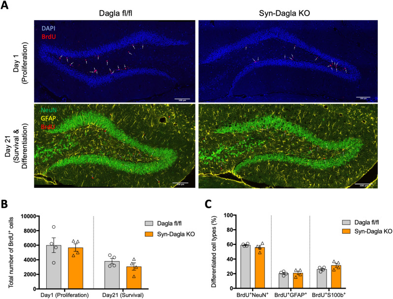
Figure 5.
Adult hippocampal neurogenesis is not altered in Syn-Dagla KO mice. (A) Representative immunohistochemistry stainings of Syn-Dagla KO and control mice. Syn-Dagla KO mice show similar number of BrdU-positive cells (red) 1 day after the last injection. 21 days after BrdU injections, the number of BrdU positive cells does not differ between Syn-Dagla KO mice and controls. Additionally, brains were stained with an astrocytic marker GFAP (yellow) and a neuronal marker NeuN (green) to investigate differentiation, which was unchanged (scale bar: 100 µm). (B) The number of BrdU-positive cells in dentate gyrus of Syn-Dagla KO mice was similar to controls, one and 21 days after BrdU injections. (C) Differentiation of progenitor cells in dentate gyrus of Syn-Dagla KO mice. BrdU-positive cells were analyzed for co-expression of neuronal marker NeuN and astrocytic markers GFAP and S100beta. Values represent mean ± SEM; n = 4 animals/group, 6 analyzed pictures/animal. Student’s t-test.

