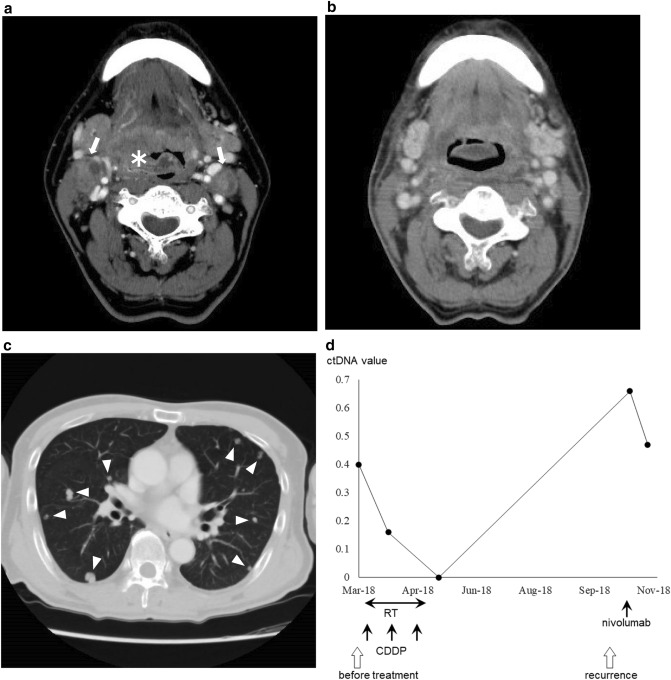
Figure 3.
(a) Pretreatment computed tomography (CT) image shows the tumor (asterisk) extending from the right lateral wall to the anterior wall of the oropharynx and cervical node metastasis (arrows). (b) CT image after initial treatment shows complete response. (c) CT image shows multiple lung metastases (triangles). (d) The pretreatment circulating tumor DNA (ctDNA) was positive and turned negative after initial treatment with concomitant chemoradiotherapy with CDDP; ctDNA was positive again at the time of local and distant recurrence and remained positive despite the administration of immune checkpoint inhibitor therapy with nivolumab.

