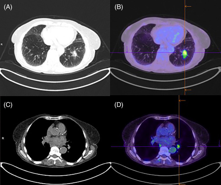
FIGURE 1.

Computed tomography/positron emission tomography (CT/PET) scan in July 2018 showing left lower lobe nodule (A) on CT, with moderate fluorodeoxyglucose (FDG) avidity (maximum standardized uptake value [SUVmax] 5.7) on PET (B), and left hilar lymphadenopathy (C) with moderate FDG avidity (SUVmax 4.3) (D)
