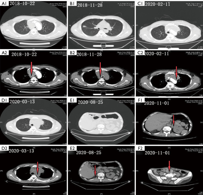
Figure 1.
Imaging results of the computed tomography (CT) can during diagnosis and treatment at different time points. (A1,A2) Chest CT showed a 16 mm × 26 mm nodule located in the right upper lobe and compressed bronchi stenosis in the inferior segment of the trachea. (B1,B2) After 1 months of Afatinib treatment, the two nodules were significantly smaller, and the efficacy was judged as a partial response. (C1,C2) After 1 months of Afatinib treatment, the efficacy was judged as progressive disease (PD). (D1,D2) Chest CT revealed right pleural effusion metastasis. (E1,E2) Chest CT showed bilateral adrenal metastasis, the efficacy was again judged as PD. (F1) The abdominal CT showed mesenchymal pelvis metastasis (F2). The red arrow represents the tumor lesion or metastasis location.

