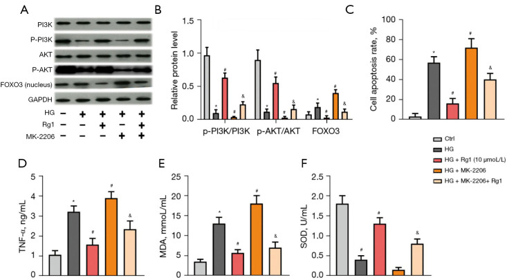
Figure 6.
The PI3K/AKT/FOXO3 pathway was involved in the protective effect of Rg1 on HG-induced HBZY-1 cells. HBZY-1 cells were induced by HG (30 mmol/L). HBZY-1 cells were divided into 5 groups: control group (HBZY-1 cells without any treatment); HG group (HBZY-1 cells were induced by 30 mmol/L glucose); HG + Rg1 (10 µmol/L) group, where HG-induced HBZY-1 cells were treated with 10 µmol/L Rg1 at 37 °C for 48 h; HG + MK-2206 (100 nmol/L) group, where HG-induced HBZY-1 cells were treated with 100 nmol/L MK-2206 at 37 °C for 48 h; HG + Rg1 + MK-2206 (100 nmol/L) group, where HG-induced HBZY-1 cells were treated with 10 µmol/L Rg1 and 100 nmol/L MK-2206 at 37 °C for 48 h. (A,B) Levels of p-PI3K/PI3K, p-AKT/AKT, and FOXO3 were measured by western blot. (C) Apoptosis rate was measured by the flow cytometry assay. (D) TNF-α level was determined by ELISA. (E) MDA level was determined by ELISA. (F) SOD activity was determined by ELISA. *P<0.05 versus control group; #P<0.05 versus the HG group; &P<0.05 versus the HG + MK-2206 (100 nmol/L) group. Rg1, ginsenoside Rg1; HG, high glucose; p-PI3K, phosphorylation of PI3K; p-AKT, phosphorylation of AKT; MDA, malondialdehyde; SOD, superoxide dismutase.

