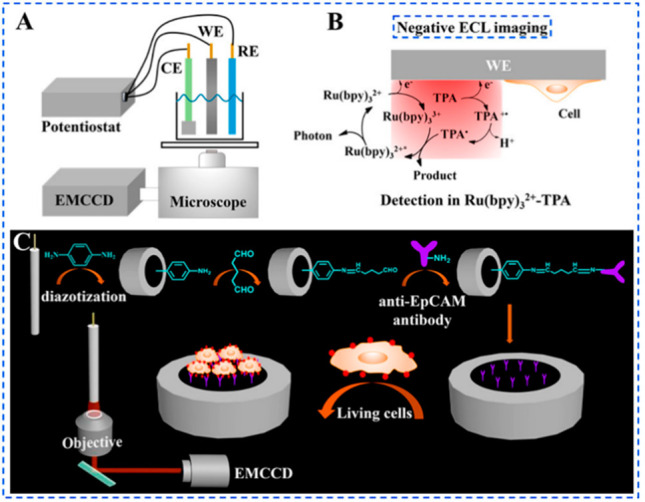
Figure 3.

(A) Instrumental setup for the negative ECL imaging system [WE, glassy carbon electrode (GCE); CE, Pt foil electrode; RE, Ag/AgCl electrode (sat KCl)]; (B) schematic illustration of the negative ECL imaging method; and (C) schematic illustration of the construction of the sensing interface and the capturing of living cells for the morphological and quantitative analyses of living cells. Reproduced from Gao, H.; Han, W.; Qi, H.; Gao, Q.; Zhang, C. Anal. Chem.2020, 92, 8278–8284 (ref (49)). Copyright 2020 American Chemical Society.
