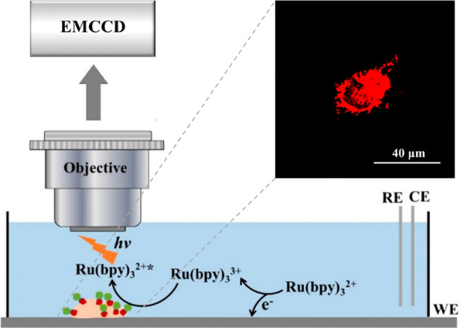
Figure 4.

ECL imaging of the carcinoembryonic antigen (CEA) on cell membranes was shown on the top-right and was detected with a microscope coupled to an EMCCD camera. Cell visualization depends on the catalytic route of ECL reactions between freely diffusing [Ru(bpy)3]2+ and Hi-AuNF@G-ssDNA-Apt coreactants. Reproduced from Chen, Y.; Gou, X.; Ma, C.; Jiang, D.; Zhu, J.-J. Anal. Chem.2021, 93, 7682–7689 (ref (63)). Copyright 2021 American Chemical Society.
