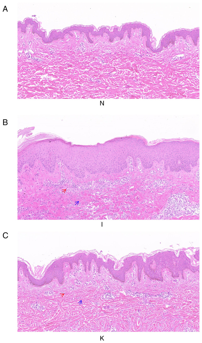
Figure 1.

Histological images of healthy skin, inflamed tissue and keloid tissue from the same patient. The three groups of samples were not consistent in morphology, representing the morphological changes of the disease from normal skin to inflammatory tissue, and finally forming keloids (A) H&E staining of group N. Magnification, ×100. (B) H&E staining of group I. Magnification, ×100. (C) H&E staining of group K. Magnification, ×100. The deep purple cells indicated by the red arrow are inflammatory cells. The blue arrows indicate collagen fibers. H&E, hematoxylin and eosin.
