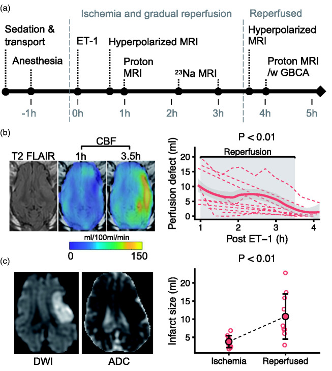
Figure 1.
Intracerebral injection of endothelin-1 (ET-1) in pigs causes ischemia and infarction followed by gradual reperfusion. After intracerebral endothelin 1 (ET-1) injection, a magnetic resonance imaging (MRI) protocol was performed including hyperpolarized MRI (a, GBCA = gadolinium-based contrast agent). Cerebral blood flow (CBF) was quantified with arterial spin labeling MRI (b). The perfusion defect (CBF < 25 ml/100 ml/min) shrank from 1 to 3.5 hours after ET-1. Diffusion weighted MRI (DWI) and apparent diffusion coefficient (ADC) maps showed a diffusion restriction 3.5 hours post ET-1 (c). The infarct (ADC < 620 × 10−6 [mm2/s]) grew from 1 hour (ischemia) to 3.5 hours (reperfused) after ET-1. Data are shown as individual observations (n = 10) with mean ± SD. In (c), a local regression curve was added for visualization (thick line). Tested with linear mixed-effect models.

