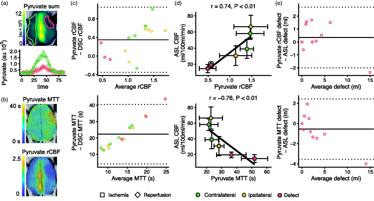
Figure 4.
Perfusion-weighted imaging with hyperpolarized [1-13C]pyruvate shows the perfusion changes following endothelin-1 induced stroke in pigs. Delivery of hyperpolarized [1-13C]pyruvate was impaired during ischemia. A large signal was observed in the sagittal sinus (a, au = arbitrary units). The time dynamics of the pyruvate signal were used to estimate perfusion via relative cerebral blood flow (rCBF) and mean transit time (MTT) following the principles of gadolinium-based dynamic susceptibility contrast (DSC) imaging (b). Bland-Altman plots showed that rCBF was slightly larger but unbiased compared with DSC, while MTT was much longer and skewed (c). The perfusion measures derived from the hyperpolarized pyruvate correlate well with CBF from arterial spin labeling MRI (ASL, d), and they showed good agreement with ASL in estimating size of the perfusion defect (e). Data examples (a and b) are from one representative animal. Infarct and contralateral brain are marked with red and green, respectively. Data are shown as individual observations (c, e) or mean ± SD (d). The curves were fitted as smooth local regressions (a) or linear regressions (d). As the same animals were examined during ischemia (n = 4) and after reperfusion (n = 7), the correlation analysis was performed under consideration of dependency in data.

