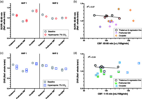
Figure 5.

(a) Tissue ratios (SUVR) relative to whole brain from the 65–80 min scan interval show almost identical values between baseline and hypercapnia conditions for each region of interest (thalamus, prefrontal gray matter (GM) and occipital cortex) in each animal. (b) Linear regressions between SUVR and cerebral blood flow (CBF) from the 65–80 scan interval do not show obvious correlations in any of the three regions (regression line shown for thalamus ROI). (c) The distribution volume ratio (DVR) shows robust outcome measurements that have similar values between baseline and hypercapnia experimental sessions. This suggests that neither outcome measurement is noticeably influenced by changes in cerebral blood flow. (d) DVR was not correlated to CBF from the 1–15 min scan interval in any of the regions, suggesting no dependency of these outcome measures on CBF.
