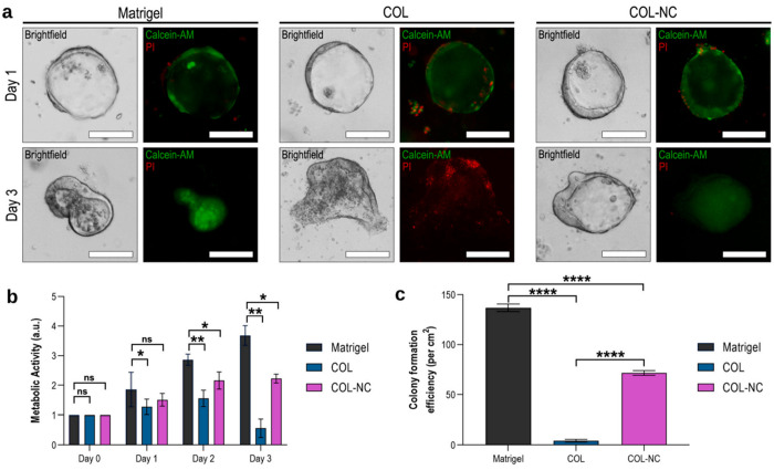
Figure 7.
Photomicrographs showing growth of intestinal organoid in various matrices. (a) Intestinal organoid grown in Matrigel, collagen (COL), and collagen–nanocellulose (COL-NC) hydrogels, as shown by bright-field and fluorescent microscopy. Organoids were stained with calcein-AM (green) and propidium iodide (PI, red) to distinguish cell viability. Live cells produce a strong green fluorescence resulting from the conversion of calcein-AM to calcein, whereas dead cells have a strong red fluorescence due to the presence of propidium iodide. (b) Metabolic activity detected in the organoids embedded in the 3 hydrogels. (c) Colony formation efficiency in three hydrogels. Reprinted with permission from ref (20). Copyright 2021 Elsevier.

