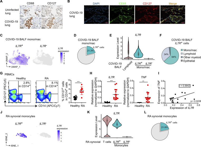
Figure 1.
CD127high monocytes/macrophages are hallmarks of human inflammatory conditions. (A and B) Immunohistochemical analysis of CD68 and CD127 expression in lung tissues sections are shown in A. Immunofluorescence staining for DAPI (blue), CD68 (green), and CD127 (red) in sections from COVID-19 lung tissues are shown in B. Uninfected lung tissues and COVID-19 lung tissues were obtained during autopsy as described in Materials and methods. One representative result from tissue sections of three COVID-19 cases is shown. Scale bar, 50 µm. (C) UMAP projection of IL7R+ and IL7R− monocytes/macrophages (mono/mac) in BALF from nine COVID-19 patients (see Materials and methods for details). IL7R expression is shown by the indicated colors. (D) Pie graph shows the percentage of IL7R+ cells in COVID-19 BALF mono/mac. (E) Violin plot shows the expression levels of IL7R in lymphoid cells and IL7R+ and IL7R− mono/mac from COVID-19 patient BALF. Each overlaid box indicates the interquartile range with median shown as a circle. (F) Pie graph shows the percentages of each cell type in total IL7R+ BALF cells from COVID-19 patients. (G) PBMCs were obtained from healthy donors (n = 13) and RA patients (n = 8), and CD127 expression was measured by FACS analysis. Representative FACS plot (left) and cumulative percentages (right) of CD127+ population in CD14+ monocytes are shown. ***, P < 0.001 by unpaired t test. Data are shown as the mean ± SD. (H) In CD14+ monocytes from PBMCs of healthy donors and RA patients, mRNA of IL7R and TNF was measured by qPCR. Relative expression was normalized to internal control (GAPDH). **, P < 0.01; ***, P < 0.001; unpaired t test. Data are shown as the mean ± SD (IL7R, healthy n = 20 and RA n = 16; TNF, healthy n = 8 and RA n = 16). (I) Linear regression analysis for the expression of TNF and IL7R in CD14+ monocytes from each RA patient in H. Correlation coefficient r and P value by F-test are labeled. (J) t-Distributed stochastic neighbor embedding (tSNE) projection of IL7R+ and IL7R− cells in RA synovial monocytes (see Materials and methods for details). IL7R expression is shown by the indicated colors. (K) Violin plot shows the expression levels of IL7R in T cells and IL7R+ and IL7R− monocytes in RA synovial tissues. Each overlaid box indicates the interquartile range with median shown as a circle. (L) Pie graph shows the percentage of IL7R+ cells in RA synovial monocytes. Each data point in G–I represents one individual donor or patient.

