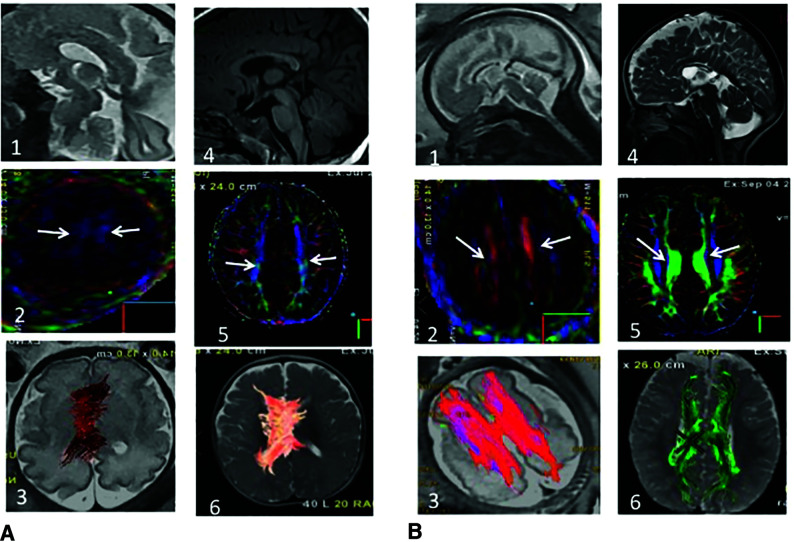
FIG 1.
Fetal (30 weeks) and postnatal anatomic MR imaging and DTI (6 months) of ISCC PB– (A) and PB+. A1, Sagittal single-shot fast spin-echo T2 (SSFSET2) shows an ISCC. A2, DTI color-coding map shows the absence of PB, confirmed in postnatal imaging (A3–6). Only the cingulum is present, well-recognized by the inferior-superior direction in blue (white arrows), present on pre- and postnatal imaging. B1, Sagittal SSFSET2 shows an ISCC. B2, DTI color-coding map shows the presence of PB (white arrows), confirmed by a postnatal color-coding map (B3–6). PB are identified by their typical anterior-posterior orientation on the color-coding maps (B2 and B5). Fiber-tracking demonstrates the typical anterior-posterior thick PB, with a remnant of the left-to-right CC (B3 and B6).

