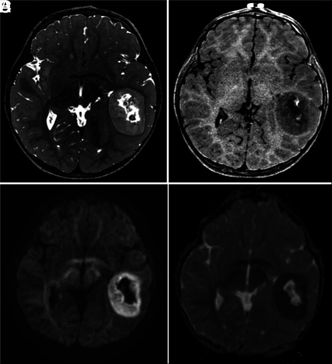
FIG 2.

Characteristic MR imaging features of CNS tumors with a BCOR ITD. A, Axial T2-weighted image shows a well-defined mass, central necrosis, and no surrounding edema. B, Axial contrast-enhanced T1-weighted image shows poor enhancement after contrast medium administration. Axial DWI (C) and ADC map (D) show restricted diffusion of the solid portions of the tumor.
