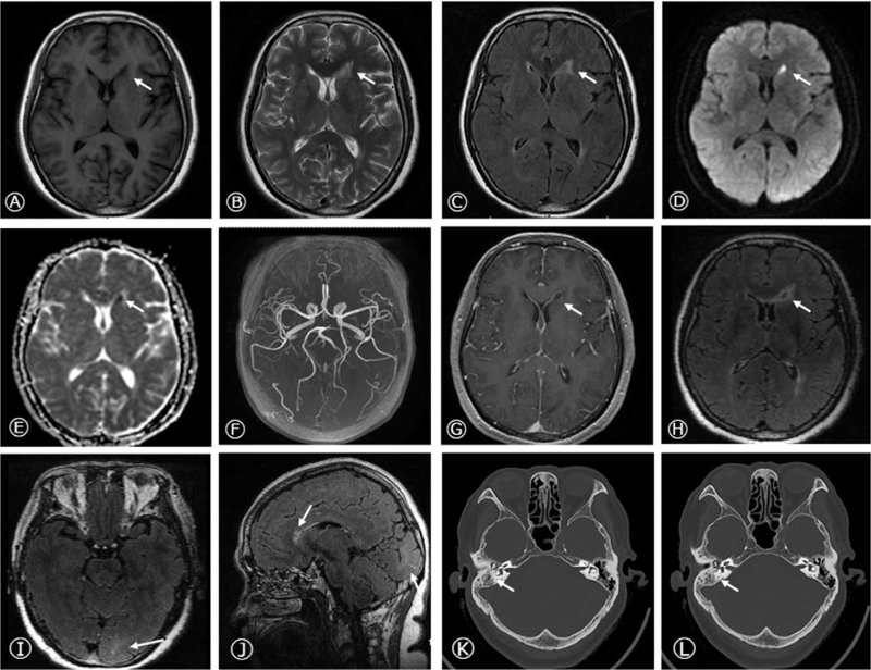
Figure 1.
Brain MRI and Temporal bone CT at admission. Axial T1-weighted sequence, T2 -weighted sequence, FLAIR sequence (A-C), DWI (D), and ADC (E) sequences were imaged and showed abnormal signals in the left head of caudate nucleus, next to the anterior horn of lateral ventricle. No significant abnormality was observed in the cerebral MRA (F). Cranial SPGR and meningeal CUBE enhancement displayed the ring-enhancement in the left head of caudate nucleus and meningeal linear enhancement in left occipital lobe on axial (G-I) and sagittal images (J). The temporal bone CT showed the right otitis mastoidea (K, L). CT = computed tomography, MRI = magnetic resonance imaging.

