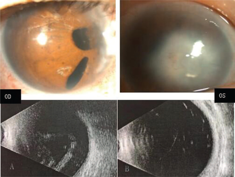
Figure 1.
Ocular characteristics of the recruited patient. Biomicroscopic photograph of the anterior segment shows iris hypoplasia with polycoria and corectopia of her right eye (A upper). Examination of the left eye shows middle corneal opacification, central iridocorneal adhesions, and peripheral corneal vascularization (B upper). Type-B ultrasonic imaging of bilateral eyes: retinal detachment in the right eye and abnormal structure of the vitreous body in the left eye (A, B lower).

