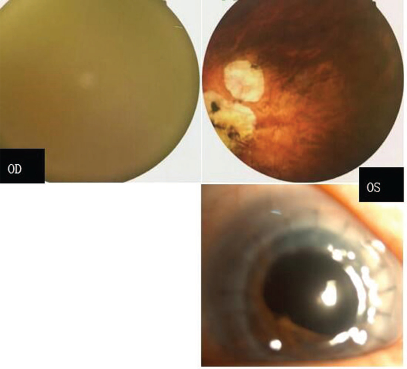
Figure 2.
The posterior segment examination of the recruited patient after the operation: the right fundus photography is not clear because of nystagmus; the left eye shows hypoplasia of the optic nerve and hypoplasia of the macula half a year after the operation (upper). The transplanted cornea is transparent (lower).

