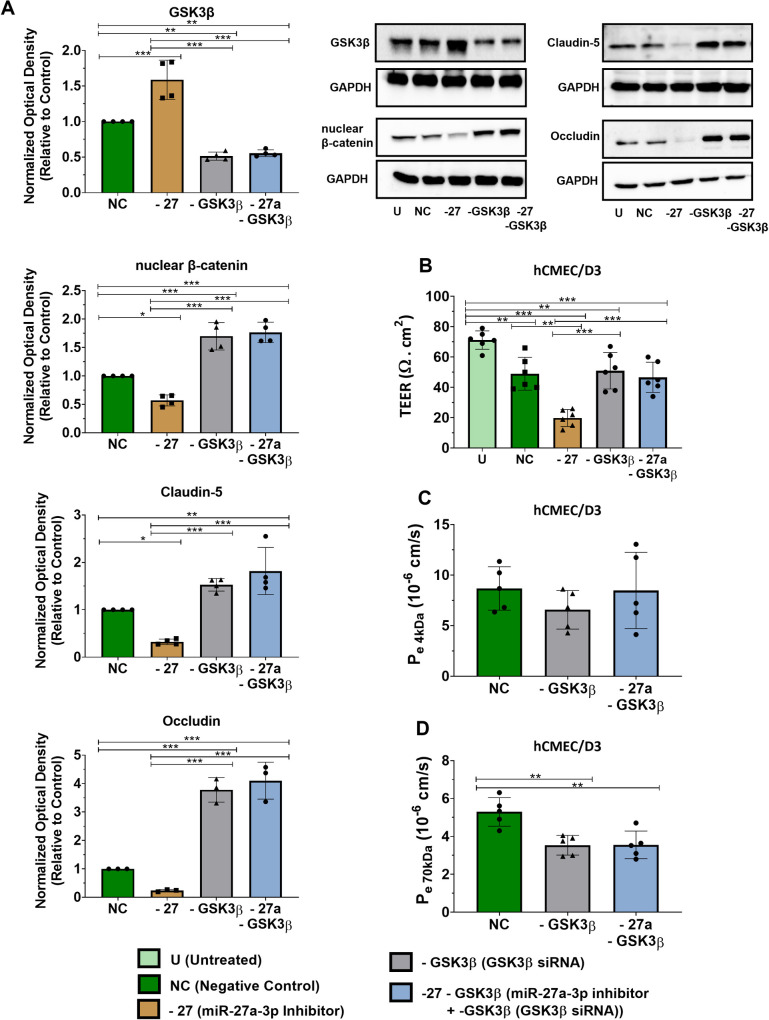
Fig 4. GSK3ß inhibition rescues the activation of GSK3ß by miR-27a-3p inhibitor.
hCMEC/D3 cells were transfected with miR-27a-3p inhibitor and/or GSK3B siRNA or negative control for 72h. (A) Protein expression of GSK3ß, nuclear ß-catenin, claudin-5 and occludin measured by western-blot in hCMEC/D3. Optical densities of three independent images were analyzed with Image Lab 6.0.1 software(Bio-Rad) and normalized to GAPDH. Results are represented as normalized optical densities. Experiments were carried out three to four times with each preparation representing pooled protein lysates from monolayer cultures performed in triplicates. (B) Transendothelial electrical resistance (TEER) of hCMEC/D3 cells treated with miR-27a-3p inhibitor and/or GSK3B siRNA or negative control after 72 hours of transfection. Experiments were carried out six times with monolayer cultures performed in triplicates. (C, D) The permeability coefficient (Pe, cm/s) of the endothelial monolayer assessed by the 4 (C) and 70 kDa FITX-dextran flux assay (D). Experiments were carried out five times with monolayer cultures performed in triplicates. Data represent mean ± SD from the independent experiments (biological replicates). *p<0.05, **p<0.01, ***p<0.001.

