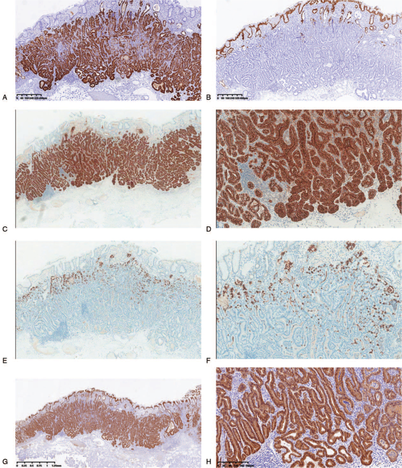
Figure 2.
Immunohistochemical staining (EnVision) results in gastric adenocarcinoma with chief cell differentiation (case 4). Carcinoma revealed diffuse positivity for MUC6 (A), but MUC5AC was only stained in the non-atypical foveolar epithelium that was covered on the surface of tumor (B). Pepsinogen-I was strongly expressed in GAFG (C and D), and focal positivity for H+/K+-ATPase (E and F). The β-catenin of all cases was only expressed in cytoplasm (G and H).

