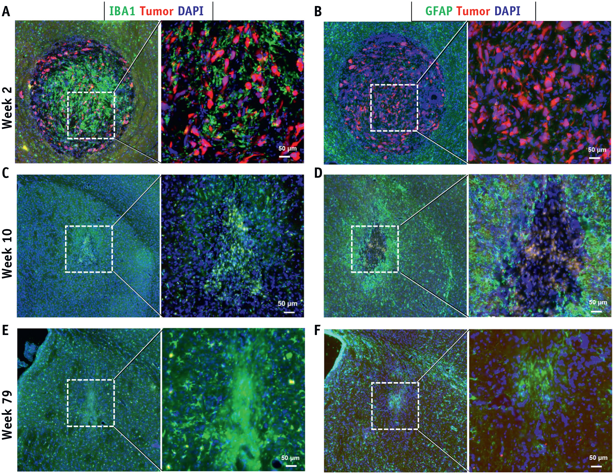
Fig. 2.

Histologic assessment of tumor response to SRS. Tumor cells were visualized by (A-F) fluorescent reporter (mCherry; Red) at weeks 2, 10, 79 after SRS. Neuroinflammation inside and surrounding the tumor core was revealed by (A, C, E) IBA1+ microglia/macrophages and (B, D, F) GFAP+ astrocytes. Abbreviation: SRS = stereotactic radiosurgery. (A color version of this figure is available at https://doi.org/10.1016/j.ijrobp.2020.05.027.)
