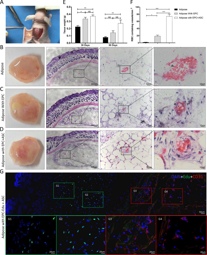
Fig 7. Coimplantation of ASCs with EPCs increases vascular density in adipose grafts.
(A) Photograph illustrating the subcutaneous transplantation of adipose tissue in nude mice. In each group, the fat implants were harvested at 30 days postimplatation and stained with HE. (B-D) Representative photographs and images of HE-stained sections of implanted adipose tissue (B), implanted adipose tissue containing EPCs (C), and implants containing a combination of ASCs and EPCs (D). Higher-magnification images of the boxed areas are shown on the right. (E) In another experiment, the fat tissue in all three groups were dissected and weighted. Data are expressed as the mean ± SEM. *, P < 0.05; **, P < 0.01; NS = not significant. (F) The density of RBC-filled vessels in the implants of each group was analyzed. Data are expressed as mean ± SEM. *, P < 0.05; **, P < 0.01. (G) Represent images of immunofluorescence staining of Edu labeled EPC and CD31 positive vessels in the adipose implants. The higher magnifications of the boxed areas are shown on the lower panel. CD31 expression was stained in red, Edu labeled EPCs was stained in green, nuclei were stained in blue with DAPI.

