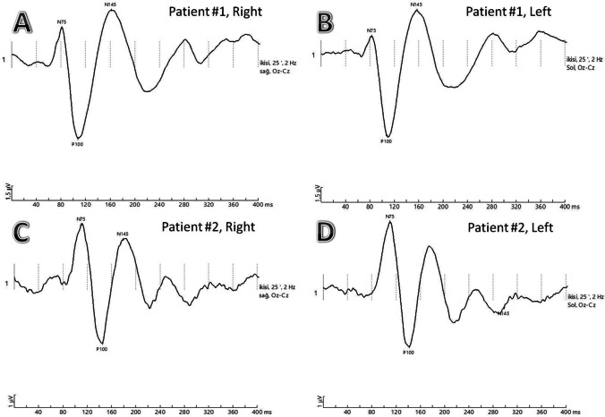
Fig. 3.
VEP recordings with N75, P100, and N145 waves are labeled A and B Patient #1, pattern reversal VEPs of a COVID-19 patient with pneumonia showing normal P100 latencies in both eyes. C and D Patient #2, pattern reversal VEPs of COVID-19 patient with pneumonia showing prolonged P100 latencies in both eyes

