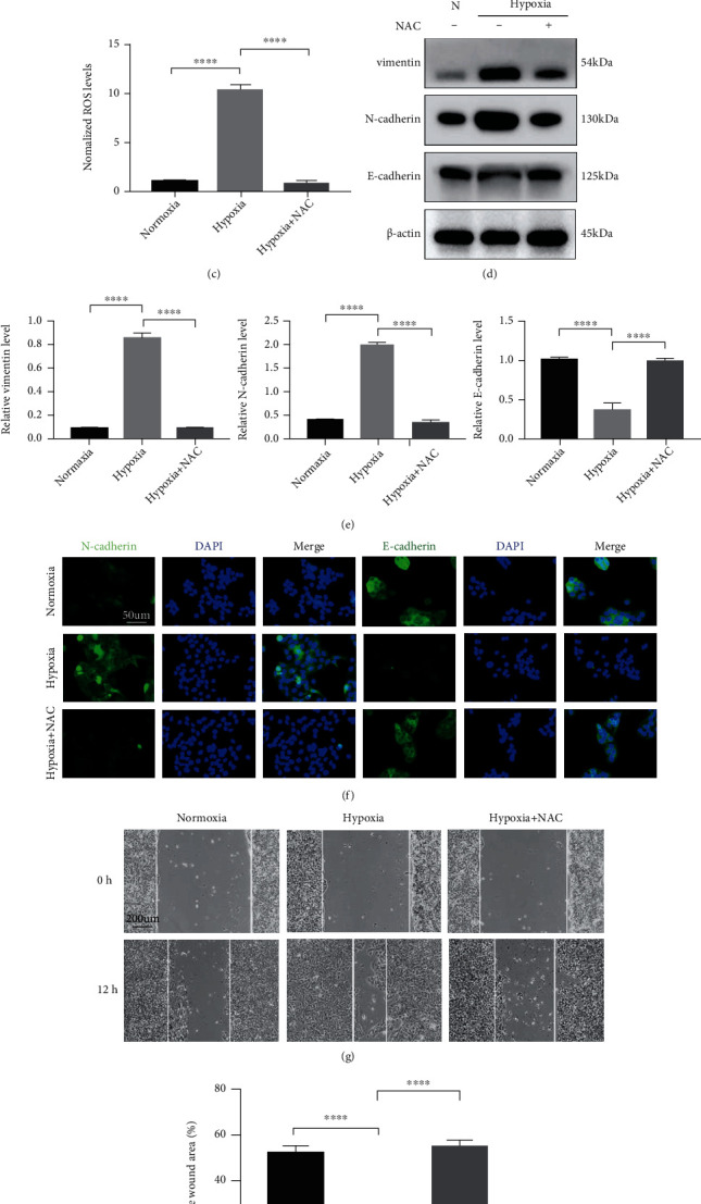
Figure 2.

ROS are required for hypoxia-stimulated EMT and migration of HaCaT cells. (a–c) Levels of endogenous ROS in HaCaT cells were measured by staining with DCFH-DA. Fluorescent DCFH-DA was monitored at the indicated time points by a microplate reader (a, c). Representative pictures showing staining with DCFH-DA (b). (d, e) Western blotting (d) and quantitative analysis (e) were performed to detect vimentin, N-cadherin, and E-cadherin levels with or without hypoxia and NAC treatment for 12 h. β-Actin was used as the loading control. (f) Representative fluorescent images of N-cadherin and E-cadherin in the indicated HaCaT cells. Bar, 50 μm. (g, h) Scratch assays (g) and quantitative analysis (h) were performed using NAC-treated and untreated HaCaT cells with or without hypoxia exposure for 12 h. Bar, 200 μm. Mean ± SEM. n = 3. ∗∗∗P < 0.001; ∗∗∗∗P < 0.0001.
