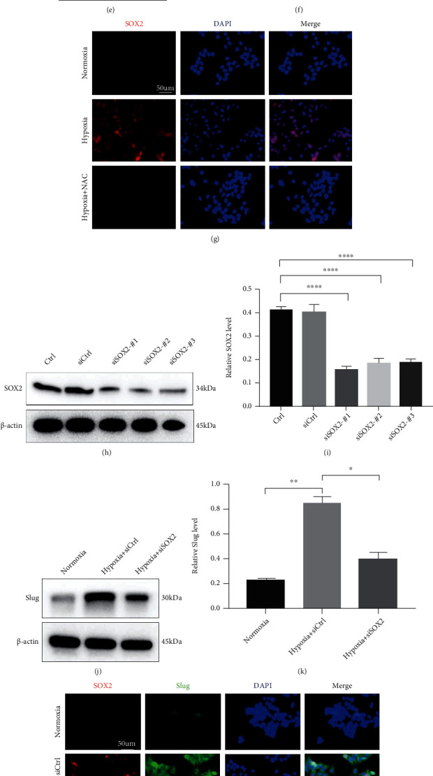
Figure 3.

The effect of SOX2 on ROS-induced HaCaT cell EMT and migration under hypoxic conditions. (a, b) HaCaT cells were incubated under hypoxia for the indicated times. SOX2 expression was assayed using Western blotting (a). Quantitative analysis (b) was employed to analyse the relative SOX2 level. SOX2 was quantified and normalized against β-actin. (c) Representative pictures of IF staining for SOX2 expression in cells treated with hypoxia or normoxia for 12 h. Bar, 50 μm. (d–f) The mRNA (d) and protein (e, f) levels of SOX2 were analysed in NAC-treated and untreated HaCaT cells with or without hypoxia treatment. (g) Representative fluorescent images of SOX2 in the indicated HaCaT cells. Bar, 50 μm. (h, i) The protein extracts from hypoxia-treated cells transfected with siSOX2 or siCtrl were analysed via Western blotting (h) and quantitative analysis (i) for SOX2. (j, k) Western blotting (j) and quantitative analysis (k) were performed to detect Slug levels in cells transfected with siSOX2 or siCtrl after 12 h of treatment with hypoxia or normoxia. (l) The expression of SOX2 (red) and Slug (green) was determined by IF staining. The representative images show typical coexpression of SOX2 and slug. Bar, 50 μm. (m, n) Scratch assays (m) and quantitative analysis (n) were performed using the indicated cells with or without hypoxia treatment for 12 h. Bar, 200 μm. Mean ± SEM. n = 3. (o) Levels of endogenous ROS were measured by a microplate reader. Mean ± SEM. n = 3. ∗P < 0.05, ∗∗P < 0.01, ∗∗∗P < 0.001, and ∗∗∗∗P < 0.0001.
