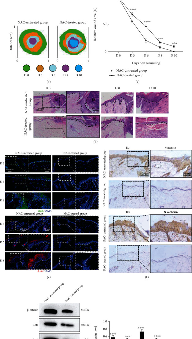
Figure 6.

ROS are required for EMT-driven reepithelialization during repair. (a–c) Representative pictures and quantitation of the healing time of skin wounds after an 8 mm primary biopsy. Biopsy sites were demarcated using blue polypropylene sutures. Bar, 2 mm. Mean ± SEM. n = 6. (d) Representative H&E-stained pictures of skin wounds on days 3, 8, and 10. The magnification of the dotted boxes is shown on the right at day 3 after wounding. Additionally, the epithelium is marked with a dotted line. Bars, 100 and 50 μm (ES: Eschar). (e) Representative pictures of wounded (days 3, 5, and 8) skin stained to show the expression of ROS (green) and SOX2 (red) in the skin (blue). The magnification of the dotted box is shown on the right of each picture. Additionally, the epithelium is marked with a dotted line. Bar, 100 and 50 μm. (f) Immunohistochemical analysis of vimentin and N-cadherin in skin wounds on day 3. The magnification of the dotted boxes is shown on the right. Bars, 100 and 50 μm. (g, h) Western blotting (g) and quantitative analysis (h) were employed to analyse the expression levels of β-catenin, Lef1, Lgr5, and Slug in the NAC-treated wounds and untreated wounds at day 3 after wounding. β-Actin was used as the loading control. Mean ± SEM. n = 3. ∗∗∗P < 0.001; ∗∗∗∗P < 0.0001.
