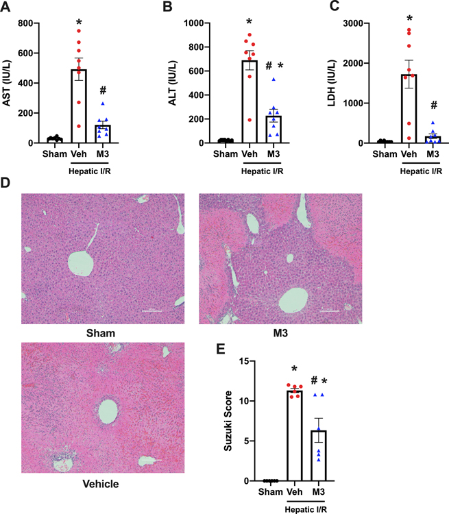
Fig. 4. M3 treatment attenuates systemic organ injury markers and decreases tissue damage after hepatic I/R.
Blood and ischemic liver tissue was collected at 24 h reperfusion from sham, vehicle (normal saline), and M3 groups. Serum (A) AST, (B) ALT, and (C) LDH were determined using specific colorimetric enzymatic assays (n = 8/group). (D), Representative images of hematoxylin & eosin stained lung tissue at 100x. Scale bar: 100 μm. (E), Extent of liver injury was graded using the Suzuki score by a blinded investigator as described in our methods (n = 6/group). Data expressed as means ± SE and compared by one-way ANOVA and SNK method (*P ≤ 0.05 versus Sham; #P ≤ 0.05 vs vehicle).

