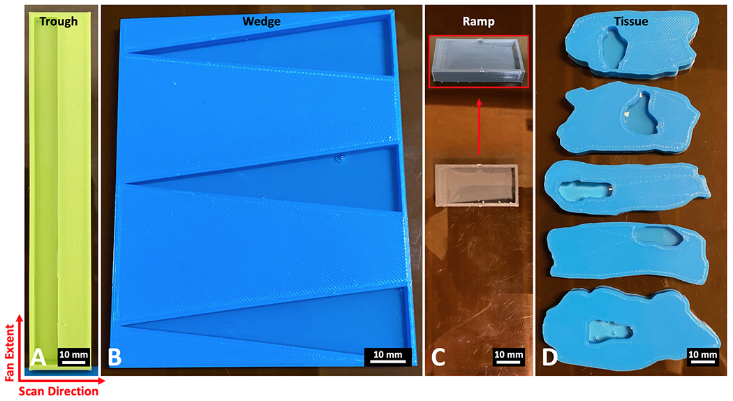
Fig 3.

Phantoms composed of water and PLA plastic (6 mm thick) for testing material classification of the X-ray fan beam coded aperture imaging system. A) A trough phantom containing rectangular regions of water and PLA B) A wedge phantom with three triangle water wells C) A ramp phantom that transitions from 6 mm PLA (left side) to 5.5 mm water (right side) by a ramp that changes the ratio of the materials through the axial dimension D) Tissue phantoms modeled after real lumpectomy tissue slices, with water wells placed at the locations of the tumors within the real samples. For imaging, the fan extends along the vertical axis of these images, while the scan direction occurs on the horizontal axis (together creating the transverse plane). The axis through the thickness of the phantoms represents the axial dimension.
