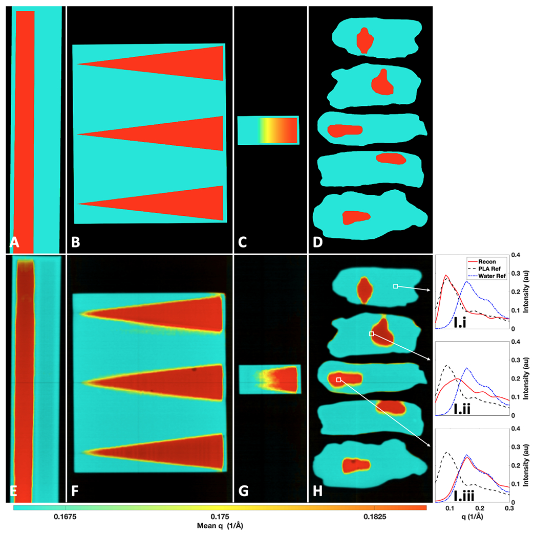
Fig 6.

Ground truth and measured X-ray transmission + momentum transfer (q) colorization (TQC) images of the water-PLA phantoms. Ground truth TQC images of the: trough (A), wedge (B), ramp (C), and tissue (D) phantoms. Measured TQC images of the: trough (E), wedge (F), ramp (G), and tissue (H) phantoms. Water and PLA correspond to red and teal coloring, respectively, on a continuum (online version only). I) The full reconstructed XRD spectra from marked regions in H containing PLA (I.i), a mixture of PLA and water (I.ii), and water (I.iii) compared against XRD spectra of water and PLA measured in a commercial diffractometer.
